A few different species of liver flukes can infect goats in North America. Fasciola hepatica, Fascioloides magna, and Dicrocoelium dendriticum (Smith and Sherman 512).
Common Liver Flukes - Fasciola Hepatica
Common liver flukes have an indirect life cycle and require aquatic snails as an intermediate host. More than 15 species of snails are known hosts. The Fasciola hepatica eggs hatch in 10-12 days at temperatures of at least 50°F and up to 79°F. The micardia (immature flukes) invade snails, where they can stay for as short of time as 21 days or up to 10 months if conditions are adverse. After leaving the snail, the immature flukes “attach to herbage, encyst, and become infective” (Smith and Sherman 515). This infective stage of fluke is called the metacercariae. The goat consumes the herbage harboring the metacercariae and is infected with the fluke.
Once in the animal, the metacercariae penetrate the liver and begin migrating through it, causing quite a lot of damage. Inflammation of the liver and bile ducts is common. Once the flukes reach the liver and mature, fluke eggs, though usually few, can be found in a fecal sedimentation test. All flukes have both male and female reproductive parts (16). Eggs are about 130-150 X 60-90 micrometers. “The entire life cycle can take a minimum of 16 weeks” (6).
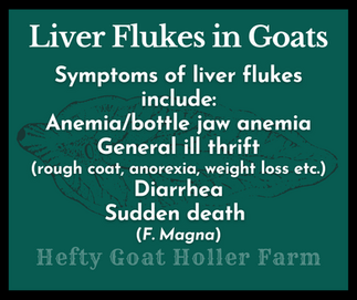
Infection can be subacute or acute, depending on the number of flukes. Feeding and movement will cause chronic blood loss in most cases. An acute infection can cause sudden death by hemorrhage but this is unusual. More likely, an acute infection would last a few days, with progressive weakness, depression, anorexia, and pallor. In subacute infections, chronic blood loss will manifest as anemia and bottle jaw. Other symptoms include depression and lethargy, poor appetite, weight loss/poor body condition, and rough hair coat. Milk production can also be negatively affected, and diarrhea is possible (4). Another source even lists constipation as a symptom since the digestive tract is just disturbed in general (6). Currently, there is little evidence goats have developed any sort of natural resistance to Fasciola hepatica infections: “Available investigations suggest that goats are highly susceptible to F. hepatica infections. Goats have a < 25% fluke recovery after F. hepatica infection, with no signs of reduced burdens upon secondary infection." (7). It is interesting to note liver flukes in sheep (the sources did not specify which species) can cause infection with Clostridium novyi type B (19) and can contribute to Red Water disease in cattle and possibly camelids due to Clostridium haemolyticum (20).
Generally speaking, these flukes require a moist environment "that allows persistent surface wetness" (Smith and Sherman 516). If aquatic snails can thrive in an area, so can common liver flukes. Pastures that are swampy and slow to drain or areas around ponds, streams, or ditches are excellent habitats. Aquatic snails can also burrow to survive dry periods. Flukes can be a year-round issue in tropical areas but are more seasonal in temperate regions, with snail populations peaking in spring and summer. This leads to the heaviest pasture contamination in late summer to early fall. In places with mild winters, late spring and early summer infections can also occur. It is interesting to note that while they do require plenty of moisture eggs also require adequate oxygen and will hatch slowly in muddy conditions.
Here is an excellent graphic illustrating the life cycle of Fasciola hepatica http://www.wormboss.com.au/cattle/worms/about-worms/worm-life-cycles-and-life-stages/fluke.php
Large American Liver Fluke - Fascioloidosis Magna
Fascioloidosis magna, also known as the large American liver fluke, has an indirect life cycle requiring aquatic snails but can be found in more species of snails covering “larger habitat, temperature, and moisture conditions” (Smith and Sherman 520). They are most common in the Great Lakes areas, the Gulf, Rockies, and Pacific Northwest (11). Sheep and goats are abnormal hosts – the preferred host of a large American liver fluke is a deer or elf. Grazing in moist areas shared with deer or elk as well as snails greatly increases the chances of sheep or goats becoming infected (17). The fluke wanders about the liver but doesn’t typically encyst since a goat is an abnormal host, meaning the producer is highly unlikely to find eggs. Symptoms might include “poor body condition and abdominal discomfort” in sheep according to Cornell University, but a single large American liver fluke can be also fatal and cause sudden death 3-6 months after initial exposure (18).
Lancet Liver Fluke - Dicrocoelium Dendriticum
The lancet liver fluke causes dicrocoeliasis, a “chronic form of liver fluke disease in goats that is less severe than that produced by Fasciola species” (Smith and Sherman 522). A goat might harbor thousands of flukes and be unaffected. Cattle, goats, and sheep are a few of the definite hosts for lancet liver flukes.
Their life cycle is different than the aforementioned species. Lancet liver flukes still require snails, but utilize terrestrial snails as an intermediate host and can be found in over 100 different species. Since they don’t require aquatic snails, their habitat is wider-spread than the common liver fluke or the Large American liver fluke.
Forested areas are a favorite of terrestrial snails and thus, potentially, lancet liver flukes. Other favored habitats include “grassy areas and near human dwellings” according to Animal Diversity.org, which lists Cionella lubrica or the glossy pillar snail as a common host (12). In theory, scientists initially believed it would be very difficult for the lancet liver fluke to expand its range since this parasite requires two specific hosts. Unfortunately, that has proven false. D. dendriticum has “a long history of colonization outside of its native host and geographical range of continental Europe” (13). Flukes were first noted in North America in the early 20th century (15). Rather recently, it was found to have spread to Alberta, Canada, residing unexpectedly in a few species of terrestrial snails there, including the subalpine mountain snail (14).
The second intermediate host is an ant, often the common black ant. After the snails ingest the lancet liver fluke eggs and the eventual asexual reproduction of various sporocysts inside the snail, the snails will cough up the developed cercariae (free-swimming larval stage) in a ball of mucous. The mucous and the slime trail left by the snail act as a protective barrier against the environment. Ants drink this slime and mucous, in turn ingesting the cercariae. Once encysted in the ant, they are called metacercariae. Amazingly, one or two metacercariae will basically turn the ant into a “zombie,” controlling its behavior at opportune times. From sunset to sunrise, the controlled ant will sit atop a blade of grass or other forage and wait to be ingested by an animal. According to “Parasitic Behaviors of the Liver Lancet Fluke” by Genevra M. Kuziel the ant “can live ‘normally’ at least a year in this condition.”
After being consumed by a grazing animal via ant, the immature flukes will travel to the bile ducts and can be found in both the liver and gall bladder. Maturation takes about 6 -7 weeks. Eggs will be produced in about 10-12 weeks. Their total life cycle takes about 6 months to complete, according to Merck Vet Manual. While in their definitive host, the lancet liver flukes will absorb nutrients through the host’s tissues. There is little data on lancet liver flukes compared to other internal parasites, but it is generally thought they are less likely to cause problems for the host than other species of flukes or that it at least takes a significant burden to produce clinical symptoms. In this study in sheep, a high burden in lambs resulted in “diarrhea and a reduced growth rate.” (1). Anemia, bottle jaw anemia, ill-thrift/weight loss, and eventual cirrhosis are possible.
Lancet liver fluke eggs can be found in a fecal float solution or sedimentation test but a float is typically more effective and certain solutions will yield better results (5). Eggs average 36-45 X 22-30 micrometers in size. For reference, a barber pole worm egg will average 70-85 X 45 micrometers. Note that these sizes will vary slightly depending on the source (9, 10).
Treating Liver Flukes
Only certain products can effectively treat flukes in goats. In the United States, these are limited to the albendazole Valbazen® and the sulfonamide clorsulon. Clorsulon is typically found in conjunction with a dewormer such as ivermectin or moxidectin. In sheep, there are studies indicating praziquantel is an effective treatment. Unfortunately, Valbazen® is only effective against mature flukes and so more than one treatment is necessary; a second is needed at approximately 21 days. There is conflicting information on the effectiveness of clorsulon in regards to fluke age. One source – Parasitpedia – lists clorsulon controls adult and “late” immature flukes but still might be needed a second or third treatment depending on the age of the flukes. Goat Medicine by Smith and Sherman lists it as “effective against all flukes two weeks of age or older.” In the case of suspected F. magna infection, “suggested treatment is albendazole at twice the recommended label dose for 2 or 3 consecutive days” (8).

The current effective dosages are reported as 7mg/kg bodyweight for clorsulon and 7.5 – 15 mg/kg bodyweight for albendazole, both administered orally. In simpler terms, this calculates to about .3cc per 10lb of clorsulon when it is a concentration of 10% or .6cc per 10lb of Valbazen® in a concentration of 11.36%.
(Special thanks to Kathy Winters for taking the time to calculate this and explain it to someone who loathes math!)
A Case of Mistaken Identity?
Liver fluke eggs, especially lancet liver fluke and common liver fluke eggs, can look quite similar to barber pole worm and other gastrointestinal parasite eggs. As mentioned in this piece, the producer will likely only see lancet liver fluke eggs since they can show up in a fecal float versus a fecal sedimentation test. Without having a way to measure the length and width of parasite eggs on a slide, it can prove difficult to distinguish between the two but lancet liver fluke eggs will be smaller than barber pole worm eggs. Lancet liver fluke eggs are operculated, meaning the ends have “lids,” while barber pole eggs do not, but these operculated ends may be hard to distinguish. The second graphic shows an operculated end. (If anyone knows the source for these two graphics, please let me know).


An Experience with Probable Dicroecoliosis
In 2018, my Kinder doe Topaz kidded with twins in late May as a first freshener. After getting over her initial terror – what WERE those loud, goop-covered things coming after her?! – she became a good dam. But I noticed her FAMACHA score was poor, and she was losing more condition than I wanted. I dewormed her in June accordingly as well as her dam, who had also kidded in May. Remember that does often experience a periparturient rise in parasites. I saw no improvement in Topaz, even with supplementing Red Cell to boost her blood-building blocks. Bindi, her dam, still had a poor FAMACHA score, though she was keeping her condition far better. A follow-up egg count on both showed the slide full of eggs! Not terribly long before this, a couple of fecal egg count reduction tests had shown my dewormer was effective, so this was very troubling and confusing. Complete resistance should not have happened that fast, especially following all the guidelines for proper deworming. I had taken Topaz off the pasture and dry-lotted her, feeding her leaves and hay, forage that would not harbor infective barber pole larvae, so I couldn't see recontamination as the cause, either.
I proceeded with a combination of dewormers – ivermectin and fenbendazole. At the time, it was recommended to use one dewormer until it was no longer effective, so a combination treatment was not my norm. I administered a copper oxide wire particle bolus, as well. Still, a follow-up fecal showed many, many eggs on the McMaster slide. During this time - a couple of weeks - Topaz was still losing weight and battling anemia that had progressed to bottle jaw anemia. Eventually, she also had loose fecals. Bindi was stable but not improving.
I was researching various things and found a post written about liver flukes. At the time, the idea that liver fluke eggs could be mistaken for barber pole worm eggs didn’t make sense to me because I hadn’t learned about LANCET liver flukes yet – I thought liver flukes were just liver flukes, that there was a single species and those eggs were not detectable in a float. Older texts and even my vet were adamant fluke infections only happened in very wet areas, but I did find a beef cattle article that showed fluke infections had occurred in as many as 26 states – that was enough to get me to think harder on the possibility of flukes.
I was desperate at this point and decided to treat both with Ivomec Plus – ivermectin and clorsulon; clorsulon is a flukicide. In just a few days, Topaz’s bottle jaw receded. With supportive care, she continued to improve. Bindi did, too. At almost 21 days on the dot after the first treatment, Topaz started developing a small submandibular swelling again. I treated her and Bindi both with a second dose of Ivomec Plus. I saw quick improvement – swelling went down within a day or two. Fecals showed no or low burdens. That was the end of it. Both got 100% better. I was left with questions that I didn’t find the answers to until later – lancet liver flukes! Below is Topaz fall of 2017, before kidding. Then shortly after kidding the following late spring (May or June?) right before I quarantined her to figure out what was going on then later that year, in October, fully recovered.
I can't say for sure those two does were experiencing a lancet liver fluke infection. But the evidence certainly leans that way to me. Nothing but a flukicide helped them. The symptoms certainly fit, as did the timeline. My herd had access to a densely wooded area where snails were thick. Our winter had been mild. Before and since then, my typical dewormer(s) have remained effective against strongyles, so I now believe what I saw on the slide were lancet liver fluke eggs, not barber pole worm eggs. I am not negating the widespread, serious issues with barber pole worms but I think, in some cases, it behooves a goat keeper to consider lancet liver flukes!
Sources:
4. Smith, M., Sherman, D. and Smith., 2009. Goat Medicine. 2nd ed. Somerset: Wiley.
8. Blackwell’s Five-Minute Veterinary Consult: Ruminant


